
Page 30 - Journal of Structural Heart Disease Volume 2, Issue 1
P. 30
Original Research Report
24
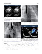
Video 26. TEE loops at 90 degrees, con rming adequate cap- ture of all margins before releasing the device from the loading cable. View supplementary video at http://dx.doi.org/10.12945/j. jshd.2016.007.14.vid.26.
Slide # 39:
Top left frame: Amplatzer septal occluder (ASO) is passed through the delivery sheath (Video 17).
Video 28.
Video 27.
Final position of the device in anteroposterior projec- tion; uroscopic “ ngerprinting” of the device. View supplementa- ry video at http://dx.doi.org/10.12945/j.jshd.2016.007.14.vid.28.
Video 29. TEE loops depicting a large ASD in a small child. The left atrium is relatively smaller compared to the right atri- um. View supplementary video at http://dx.doi.org/10.12945/j. jshd.2016.007.14.vid.29.
Top right frame: Left atrial disk of the ASO is ex- truded in the left atrium (Video 18).
Bottom frame: Corresponding TEE loop showing left atrial disk in the left atrium (Video 19).
Release of the ASO from loading cable in left anterior oblique (LAO) view. Note the well-separated disks of the ASO in LAO view con rming a well-positioned device. View supplemen- tary video at http://dx.doi.org/10.12945/j.jshd.2016.007.14.vid.27.
Journal of Structural Heart Disease, February 2016
Volume 2, Issue 1:15-32
