
Page 34 - Journal of Structural Heart Disease Volume 2, Issue 1
P. 34
Original Research Report
28
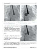
Video 40. An Occlutech balloon is positioned in the right atri- um and pushed against the interatrial septum over a Supersti wire positioned in the LSPV. View supplementary video at http:// dx.doi.org/10.12945/j.jshd.2016.007.14.vid.40.
Middle frame: ASO is deployed with the left atrial disk engaged into the right superior pulmonary vein. Loading cable is pushed to disengage the let atrial disk from the RSPV (Video 36).
Right frame: ASO is deployed with the left atrial disk engaged in the left atrial appendage. Similar to the previous case, the LA disk has been disengaged from the LA appendage by pushing the loading cable (Video 37).
Slide # 51:
Left frame: The delivery sheath has been posi- tioned outside the right superior pulmonary vein in- stead of LSPV to prevent malalignment of the device disks with the interatrial septum (Video 38).
Right frame: The ASO device is deployed by this technique (Video 39).
Slide # 52:
Left frame: Hausdorf sheath (Cook, Bloomington, Indiana, USA) is a specially designed long sheath with two posterior curves at its end, allowing for a
Video 41. The balloon is in ated followed by sequential release of the left atrial disk, waist and the right atrial disk. Supersti wire positioned in the LSPV. View supplementary video at http://dx. doi.org/10.12945/j.jshd.2016.007.14.vid.41.
Video 42. The balloon is gradually de ated to allow de- ployment of the device across the ASD. Supersti wire posi- tioned in the LSPV. View supplementary video at http://dx.doi. org/10.12945/j.jshd.2016.007.14.vid.42.
Journal of Structural Heart Disease, February 2016
Volume 2, Issue 1:15-32
