
Page 33 - Journal of Structural Heart Disease Volume 2, Issue 1
P. 33
27
Original Research Report
Video 37. ASO is deployed with the left atrial disk engaged in the left atrial appendage. Similar to the previous case, the LA disk has been disengaged from the LA appendage by pushing the loading cable. View supplementary video at http://dx.doi. org/10.12945/j.jshd.2016.007.14.vid.37.
Slide # 48:
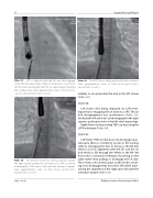
Left frame: ASO being deployed via LSPV tech- nique that is engaging the LA disk into LSPV. The LA disk disengagement was spontaneous (Video 33). Similarly the left atrial disc can be engaged in the right superior pulmonary vein or the left atrial appendage.
Right frame: Corresponding TEE loop depicting the LSPV technique (Video 34).
Slide # 49:
Left frame: If the LA disk does not disengage spon- taneously, there is a tendency to pull on the loading cable to disengage the disk. In doing so, the RA disk tends to lose its alignment with the IAS and the LA disk tends to fall through the defect in the RA. We have used a contrarian technique of pushing on the cable rather than pulling, to disengage the LA disk. This creates a secondary torque on the LA disc, result- ing in its disengagement from the LSPV while main- taining the alignment of the right atrial disk with the interatrial septum (Video 35).
The delivery sheath has been positioned outside the right superior pulmonary vein instead of LSPV to prevent malalignment of the device disks with the interatrial septum. View supplementary video at http://dx.doi.org/10.12945/j. jshd.2016.007.14.vid.38.
Video 39. The ASO device being deployed by this technique. View supplementary video at http://dx.doi.org/10.12945/j. jshd.2016.007.14.vid.39.
inability to accommodate the disk in the left atrium (Video 32).
Video 38.
Jain, S. et al.
Catheter closure of atrial septal defect
