Original Research Articles
 Download PDF (6.8 MB)
Journal of Structural Heart Disease, May 2015, Volume 1, Issue 1:9-19
DOI: 10.12945/j.jshd.2015.00011-14
Real-Time 3D Transesophageal Echocardiographic Guidance of Prosthetic Valve Paravalvular Leak
Joseph M. Venturini MD, Anuj Mediratta MD, Karima Addetia MD, Sandeep Nathan MD, Atman P. Shah MD, Roberto M. Lang MD
University of Chicago Medical Center, Section of Cardiology, Chicago, Illinois, USA
The first two authors contributed equally to the manuscript.
Abstract
Paravalvular leak (PVL), defined as retrograde blood flow adjacent to an annuloplasty ring or prosthetic valve, is a rare but serious complication of heart valve surgery. Though most PVLs are asymptomatic, 1–5% of patients develop serious clinical consequences such as heart failure, endocarditis, or hemolysis. Surgical repair may be necessary in severe cases, however for those who are at high surgical risk, a percutaneous approach can be performed to occlude these defects. Real-time three-dimensional transesophogeal echocardiography (3DTEE) during percutaneous closure procedures is invaluable for intra-procedural guidance. In this article, we will review the literature and outline two cases where real-time 3DTEE guidance was critical for successful closure of symptomatic PVL.
Video 1
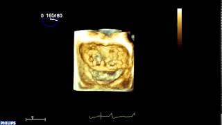
Video 1: Annuloplasty ring dehiscence.
Video 2
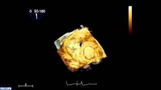
Video 2: Mechanical mitral prosthetic dehiscence.
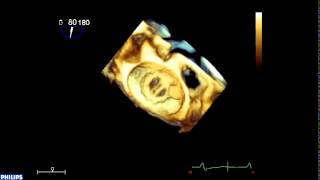
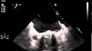
Video 3

Video 3: Case 1: Mechanical mitral prosthesis with paravalvular leak in the 4 o’clock position.
Video 4
Video 4: Case 1: 3D acquisition of 6 mm occluder device expansion.
Video 5
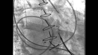
Video 5: Case 2: Fluoroscopy showing evidence of entrapment of the posterior mechanical mitral valve leaflet.
Video 6
Video 6: Case 2: 2D TEE showing evidence of entrapment of the posterior mechanical mitral valve leaflet.
Video 7
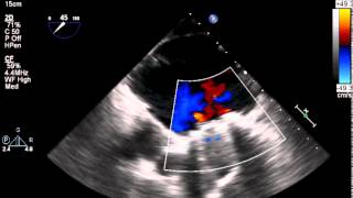
Video 7: Case 2: 2D color Doppler TEE showing evidence of entrapment of the posterior mechanical mitral valve leaflet.
Cite this article as: Venturini JM, Mediratta A, Addetia K, Nathan S, Shah AP, Lang RM. Real-Time 3D Transesophageal Echocardiographic Guidance of Prosthetic Valve Paravalvular Leak. Structural Heart Disease 2015;1(1):9-19. DOI: 10.12945/j.jshd.2015.00011-14
All comments will be screened and reviewed before posting. Statements, opinions, and results of studies published in Journal of Structural Heart Disease are those of the authors and do not reflect the policy or position of The Journal and Science International and the Editorial Board and provides no warranty as to their accuracy or reliability. Material is copyrighted and owned by Science International and cannot be used without expressed permission.
Ziyad Hijazi — May 17, 2016 7:11 PM
Sidra Medical & Research Center — Pediatrics —
San Diego, CA United States
Original Research Articles
Journal of Structural Heart Disease, May 2015, Volume 1, Issue 1:9-19
DOI: 10.12945/j.jshd.2015.00011-14
Real-Time 3D Transesophageal Echocardiographic Guidance of Prosthetic Valve Paravalvular Leak
Joseph M. Venturini MD, Anuj Mediratta MD, Karima Addetia MD, Sandeep Nathan MD, Atman P. Shah MD, Roberto M. Lang MD
University of Chicago Medical Center, Section of Cardiology, Chicago, Illinois, USA
The first two authors contributed equally to the manuscript.
Abstract
Paravalvular leak (PVL), defined as retrograde blood flow adjacent to an annuloplasty ring or prosthetic valve, is a rare but serious complication of heart valve surgery. Though most PVLs are asymptomatic, 1–5% of patients develop serious clinical consequences such as heart failure, endocarditis, or hemolysis. Surgical repair may be necessary in severe cases, however for those who are at high surgical risk, a percutaneous approach can be performed to occlude these defects. Real-time three-dimensional transesophogeal echocardiography (3DTEE) during percutaneous closure procedures is invaluable for intra-procedural guidance. In this article, we will review the literature and outline two cases where real-time 3DTEE guidance was critical for successful closure of symptomatic PVL.
Supplemental Media
Video 1
Video 2
Video 3
Video 4
Video 5
Video 6
Video 7
PDF
Mobile-ready Flipbook
Cite this article as: Venturini JM, Mediratta A, Addetia K, Nathan S, Shah AP, Lang RM. Real-Time 3D Transesophageal Echocardiographic Guidance of Prosthetic Valve Paravalvular Leak. Structural Heart Disease 2015;1(1):9-19. DOI: 10.12945/j.jshd.2015.00011-14
You must be registered and logged in to leave comments.
Ask a question (publicly)
Sidra Medical & Research Center — Pediatrics — San Diego, CA United States